Jpn. J. Infect. Dis., 207-210, 2002
To see a printable version of the article in the Adobe file format, click this [PDF] link.
Laboratory and Epidemiology Communications
Phylogenetic Analysis of Salmonella enterica Serovar Enteritidis Isolates from Food Poisoning Outbreaks and Sporadic Infections in 2001-2002 in Hyogo Prefecture: Existence of Predominant Genotypes in the Epidemic
Koichi Hamada*, Hidetaka Tsuji, Tomohiro Oshibe and Kohori Oshima
Infectious Disease Research Division, Hyogo Prefectural Institute of Public Health and Environmental Sciences, Arata-cho 2-1-29, Hyogo-ku, Kobe 652-0032
Communicated by Takashi Kawamura
Since 1989, Salmonella enterica serovar Enteritidis has become the most prevalent among the Salmonella isolates in Japan (1). We previously analyzed, by means of phylogenetic dendrograms of pulsed-field gel electrophoresis (PFGE), genetic variations of the Salmonella isolates from 28 food poisonings in 1997-2000 (2,3) and 55 cases of sporadic infections in 1996-2000 (4). In the present study, we analyzed isolates from 15 food poisonings and 12 sporadically infected cases in 2001-2002 and compared the results with those of the above cases in 1996-2000. We found predominant genotypes throughout the epidemics that took place over the past several years in our prefecture.
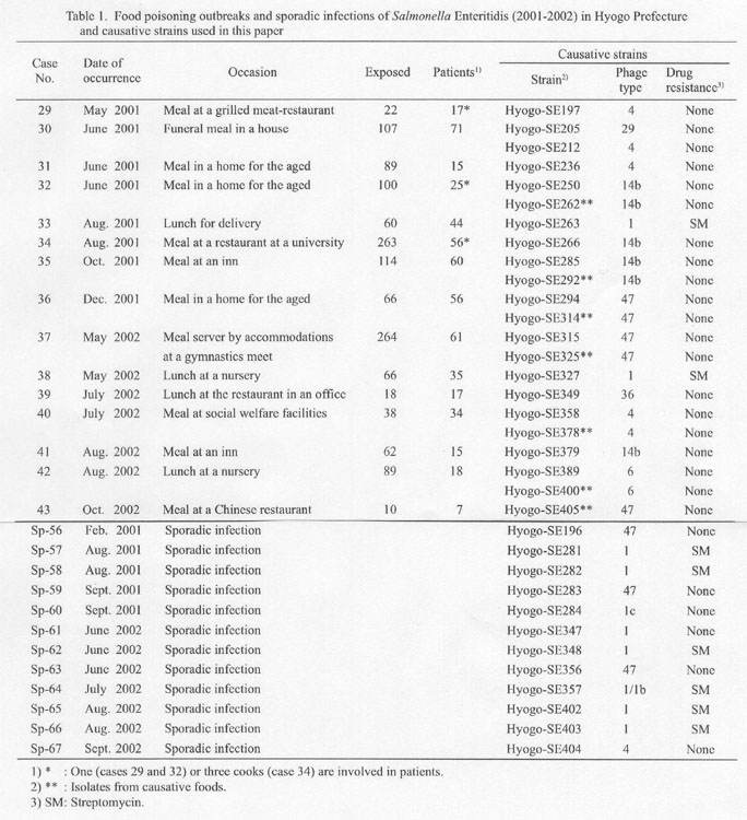
We analyzed a total of 34 specimens; 22 isolates in 15 food poisoning outbreaks (15 from stool specimens and 7 from suspected foods) and 12 isolates (all from stool specimens) in 12 sporadic infections (Table 1). The identified phage types (PTs) pf these isolates were PT1, PT1/1b, PTlc, PT4, PT6, PT14b, PT29, PT36, and PT47 (Table 1). As in previous cases (2-4), there was no correlation between PTs and PFGE aptterns (see Table 1 and Figs. 1 and 2). Antibiotice sensitivity test using antibiotic disks (Becton Dickinson Microbiology Systems, Cockeysville, Md., USA) (5) revealed that all the isolates were sensitive to ampicillin, cefotaxime, kanamycin, gentamicin, streptomycin (SM), tetracycline, trimethoprim, ciplofloxacin, fosfomycin, chloramphenicol, sulphamethoxazole-trimethoprim, and nalidixic acid except eight silates sensitive to all but SM (Table 1).
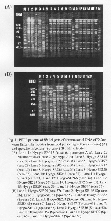
We analyzed the PFGE patterns of the isolates employing a Gene Path Typing System (Program No. 2; Bio-Rad Laboratories, Hercules, Calif., USA). As shown in Fig. 1, the PFGE patterns of bacterial chromosomal DNAs digested with BlnI (Takara Shuzo, Co., Ltd., Kyoto) resembled the A types, A-a and A-b, observed in the past (3). A-a and A-b were quite similar; their only difference was that a >630 kb band in A-a (see Hyogo-SE017, lane 1 in Fig. 1A) migrated slightly more slowly that that in A-b (see Nihinomiya-01, lane 2 in Fig. 1A). In 15 food poisoning cases (Fig. 1A), 13 were caused by A types. Only two, Hyogo-SE327 from case 38 (lane 4) and Hyogo-SE263 from case 33 (lane 11) were different. In contrast, among 12 sporadic cases (Fig. 1B), six cases, Sp-cases 57, 58, 62, 64, 65, and 66 (lanes 3, 4, 8, 10, 11, and 12, respectively) were not A types.
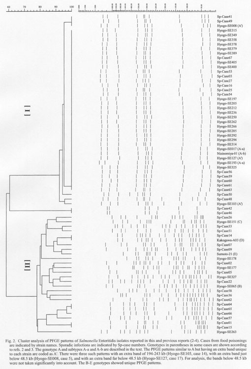
A cluster analysis (Finger Printing Plus; Bio-Rad) of the present isolates and those in the past years, 1996-2000 (2-4), together (Fig. 2) indicated presence of two large clusters with low similarity (approximately 60%): a main cluster (cluster I) including A-a, A b, and A' (2) types (see the footnote in Fig. 2) and a minor one (cluster II) constituted of unique genotypes such as B-E types (2) (see the footnote in Fig. 2). The cluster I was homegeneous except two isolates, Sp-case 42 and Sp-case 46, that probably constitute a sub-cluster. Cluster II consisted of several sub-clusters.
Our data clearly showed that most food poisoning outbreaks and half of the sporadic cases in 1996-2002 were caused by S. enterica serovar Enteritidis whose genotype belongs to cluster I. This observation is largely different from that regarding the enterohemorragic Escherichia coli O157 epidemic in Japan since 1996, which rarely showed predominant genotypes throughout the epidemic (6). There was no correlation between PTs and PFGE patterns in the S. Enteritidis epidemic as indicated here and previously (2-4).
The authors are grateful to Drs. Hidemasa Izumiya and Haruo Watanabe, National Institute of Infectious Diseases, Tokyo, for the phage typing of our isolates and for their valuable comments and advice regading this work.
References
*Corresponding author: Tel: +81-78-511-6787, Fax: +81-78-531-7080
Go to JJID Homepage Go to JJID Contents 55 (6)