Echinococcosis in Japan, 1999-2018
(IASR Vol. 40 p33-34: March, 2019)
Echinococcosis is a zoonotic helminthiasis caused by cestodes of the genus Echinococcus. The life cycle of Echinococcus involves two host species: the definitive host, in which the adult tapeworms parasitize and undergo sexual reproduction, and the intermediate host that larvae infest and undergo asexual reproduction. In general, humans are not involved in transmission, but like the intermediate hosts, humans are infected by ingestion of eggs excreted by the definitive hosts. Larvae parasitized in parenchymal organs (mainly the liver) repeat proliferation and metastasis, leading to a fatal course unless treated appropriately. There are several species of the genus Echinococcus, but regarding public health, the important species are E. multilocularis, which causes alveolar echinococcosis (with domestic distribution), and E. granulosus, which causes cystic echinococcosis (without domestic distribution). Echinococcosis caused by both species is a Category Ⅳ infectious disease according to the Infectious Diseases Control Law, which took effect in April 1999, and physicians who diagnose echinococcosis must immediately notify the public health center with jurisdiction. In addition, due to the enactment of the revised Infectious Diseases Control Law in October 2004, notification by veterinarians who diagnose echinococcosis in dogs, which can be a source of transmission, was mandated.
Human echinococcosis
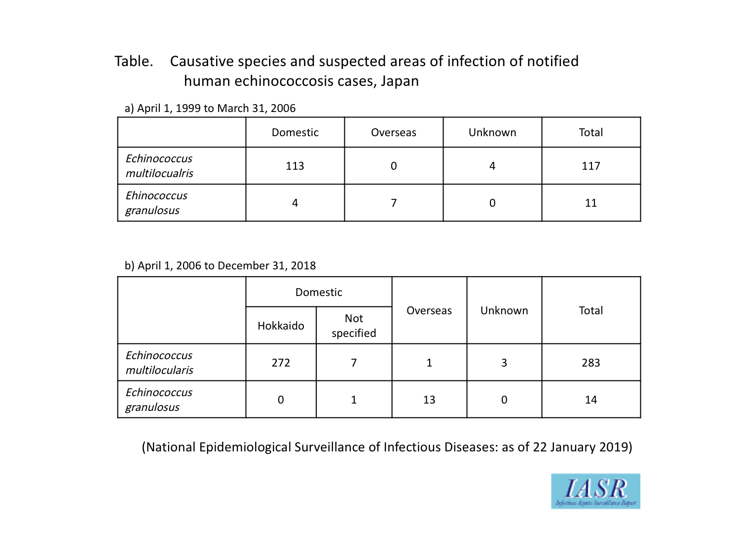
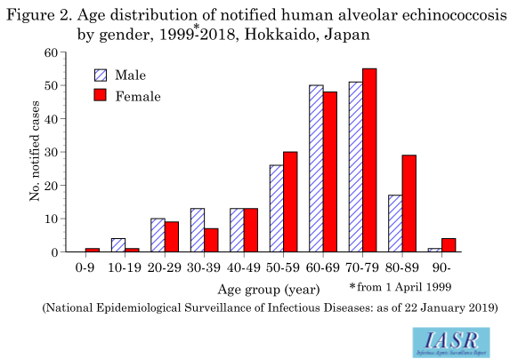
Number of cases: The notification status of human echinococcosis to the National Epidemiological Surveillance of Infectious Diseases (NESID) system conducted under the Infectious Diseases Control Law is shown in Figure 1. From April 1999 to the end of 2018 (as of January 22, 2019), 425 cases were notified, with 400 cases (94%) according to the notification forms being alveolar echinococcosis, and more than 95% of these cases were from Hokkaido, a domestic endemic area (382 cases). The suspected areas of infection were 392 cases in Japan, 1 overseas, and 7 unknown (Table). As for the gender age distribution of cases notified from Hokkaido, the sex ratio was 1:1.1, the median age was 65 years (64 years for males, 67 years for females), and a peak was noted in elderly populations (Figure 2). Outside Hokkaido, 18 cases were notified from 10 prefectures, with 5 cases from Tokyo, 2 cases each from Aomori, Kanagawa and Aichi, and 1 case each from Yamagata, Saitama, Chiba, Fukui, Mie, Osaka and Yamaguchi. On the other hand, 15 out of 25 cases (60%) notified as cystic echinococcosis were estimated to be in foreigners or those of Japanese ancestry living in Japan based on the names and remarks in the notification form, and the suspected areas of infection were 4 cases each from China and Peru, 2 cases each from Afghanistan, Nepal and Pakistan, and 1 case each from Iran, Uzbekistan and Syria (overlaps exist). Half of the 10 cases of cystic echinococcosis in Japanese individuals had a history of having been in an area where the disease was known to be endemic. Of the remaining half, 4 cases had suspected areas of infection in Japan, and the place of infection in one case was not specified (Table). The 4 cases of domestic infection were notified from Hokkaido, although it is unknown whether the actual causative agent was E. granulosus.
Clinical presentation: The growth of cysts that form in organs is slow, and the clinical course progresses without any symptoms while the cysts are small. For this reason, it is difficult to identify the infection without screening tests. Subjective symptoms appear after a prolonged period (several years to more than 10 years). In the 297 cases notified after April 2006, when the NESID notification forms began including options in the “Symptoms” column to indicate symptoms, “Abnormal diagnostic imaging findings in the liver” was the most common and observed in 208 cases (70%), followed by “Others (liver dysfunction etc.)” in 56 cases (19%), “hepatomegaly” in 38 cases (13%) and “abdominal pain” in 37 cases (12%) (overlaps exist) (see pp. 35 & 36 of this issue).
Testing and diagnosis: According to the NESID notification forms for 425 cases, the diagnosis was performed by diagnostic imaging in 237 cases (56%) and by serological diagnosis using ELISA or Western Blotting in 336 cases (79%) (overlaps exist) (see p. 38 of this issue). Pathogen detection using biopsy samples or surgical material was performed in 157 cases (37%) (overlaps exist). Although serological diagnosis of echinococcosis can be outsourced to commercial laboratories, there have been sporadic cases in which cystic echinococcosis was examined using the E. multilocularis antigen and judged as “negative”. Upon request, it is necessary to confirm the examination procedure in detail. The National Institute of Infectious Diseases will assist physicians if cystic echinococcosis is suspected.
Treatment: In the case of alveolar echinococcosis, complete resection of the lesion is the only radical treatment; therefore, early diagnosis before the invasion and expansion of the lesion is emphasized. Drug treatment using albendazole is performed in cases with lesion remnants or unresectable cases, but the effects are not constant. Development of novel chemotherapeutic agents focusing on parasite-specific energy metabolism is now underway (see p. 39 of this issue). In cystic echinococcosis where isolated lesions are common, surgical resection of cysts, or treatment by PAIR (Puncture, Aspiration, Injection, Re-aspiration) or administration of albendazole is usually performed, and an improved prognosis can be expected as compared with alveolar echinococcosis (see pp. 35 & 36 of this issue).
Canine echinococcosis
Number of cases: The number of notified cases to the NESID system until 2018 was 22 (Figure 3). The increase in the number of notifications in 2018 was due to the detection of infectious cases in epidemiological studies. Although it is not described in the NESID notification forms, all cases are believed to have been due to E. multilocularis. Seventeen cases were notified from Hokkaido, whereas 5 cases were notified from prefectures outside Hokkaido (1 case from Saitama, 4 cases from Aichi). Of the 22 cases notified, 16 cases were identified in animal hospitals (all cases in Hokkaido), whereas the remaining 6 cases were from dogs in public animal shelters (1 case in Hokkaido, 5 cases outside Hokkaido) (see p. 40 of this issue).
Clinical presentation: As the adult Echinococcus attaches to the small intestine mucosa of the definitive host (dogs and foxes) using hooks and suckers, bloody mucoid stool and watery diarrhea may develop (particularly during the early stages of infection), if a large number of worms parasitize the intestines, although the conditions are generally asymptomatic. Of the 22 cases notified, 4 cases had some type of gastrointestinal symptom.
Testing and diagnosis: Among the 22 cases notified to the NESID system, gene detection by PCR using isolated eggs or whole stool samples was performed in 17 cases, coproantigen detection by ELISA was performed in 10 cases and microscopic egg detection was performed in 8 cases (overlaps exist). As the morphological characteristics of Echinococcus eggs are indistinguishable from those of closely related taeniid species, molecular identification by PCR amplification is essential when eggs are detected. Furthermore, as ELISA can generate false positives due to non-specific reactions, re-examination must be conducted after administering anthelmintic drugs to confirm negative conversion of the result.
Treatment: Praziquantel is used to treat the definitive host. If there is no mixed infection with other kinds of cestodes, 100% deworming effects can be expected with the standard dose. However, as praziquantel has no ovicidal effects, and feces after administration can be a source of infection to humans, sufficient care must be taken when disposing feces (see p. 42 of this issue).
Prevention and countermeasures for echinococcosis
EG95, a recombinant antigen from the E. granulosus eggs, has been developed as a vaccine targeting intermediate hosts, including humans, and trials using sheep have been conducted. However, it has not been put to practical use. Therefore, not ingesting the parasite eggs is the only means of prevention, and in Hokkaido, countermeasures are being taken assuming different routes of infection (see p. 43 of this issue). In order to reduce the number of parasite eggs in the living environment, distribution of bait containing anthelmintics for wild foxes has been attempted (see p. 45 of this issue), and prophylactic administration of anthelmintic drugs to dogs is also being considered. Development of definitive host vaccines has also started (see p. 42 of this issue). In addition to these efforts, screening of residents of Hokkaido by antibody tests has been conducted from the viewpoint of early detection and prompt treatment, and improvemts have been made to increase the accuracy of the tests (see p. 38 of this issue).
When echinococcosis was last featured in the IASR (IASR 20: 1-2, 1999), transmission to Honshu, the main island of Japan, was already a concern, but indigenously infected animals were not identified. Today, however, infected dogs are continuously found in a part of Aichi Prefecture, and it should be noted that infection sources may also exist outside Hokkaido. Although echinococcosis is still rare in prefectures south of Honshu, efforts should be made to obtain more detailed patient information, such as residential history and travel history, to pursue the source of infection of the patient, and it is also necessary to establish a nationwide surveillance system for potential host animals (see p. 40 of this issue).