Mycoses in Japan as of 2012
(IASR 34: 1-2, January 2013)
Mycoses and responsible pathogens
Mycoses, diseases caused by pathogenic fungi, are classified into two categories depending upon the site of lesion: “superficial mycosis” affecting epidermal or mucosal tissues and “systemic mycosis” producing pathological changes which are disseminated in the whole body or localized to some internal organs. Though allergenic diseases or poisoning caused by fungi (e.g., aflatoxin) are sometimes included in the category of mycosis, this article principally deals with systemic mycosis, which is often fatal.
Fungi causing systemic mycosis are largely divided into two categories, those causing diseases among normal population, such as, genus Coccidioides, genus Histoplasma and genus Cryptococcus, and those causing diseases only among immunodeficient people, such as, genus Candida and genus Aspergillus. Among systemic mycosis, cryptococcosis is found in Japan, but coccidioidosis, histoplasmosis and some other mycoses, such as paracoccidioidosis, can be acquired only abroad, which are categorized as imported mycoses (IASR 23: 55-56, 2002).
Coccidioidomycosis is a category IV infectious disease in the scheme of National Epidemiological Surveillance of Infectious Diseases under the Law Concerning the Prevention of Infectious Diseases and Medical Care for Patients of Infections (Infectious Diseases Control Law, in short), which obliges doctors who made diagnosis of this infection to notify every case (See http://www.mhlw.go.jp/bunya/kenkou/kekkaku-kansenshou11/01-04-12.html for criteria of notification).
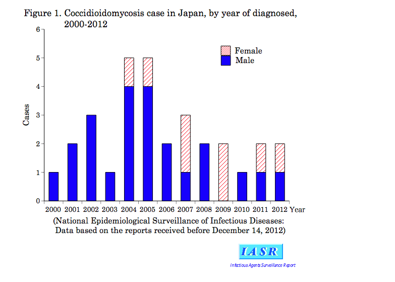
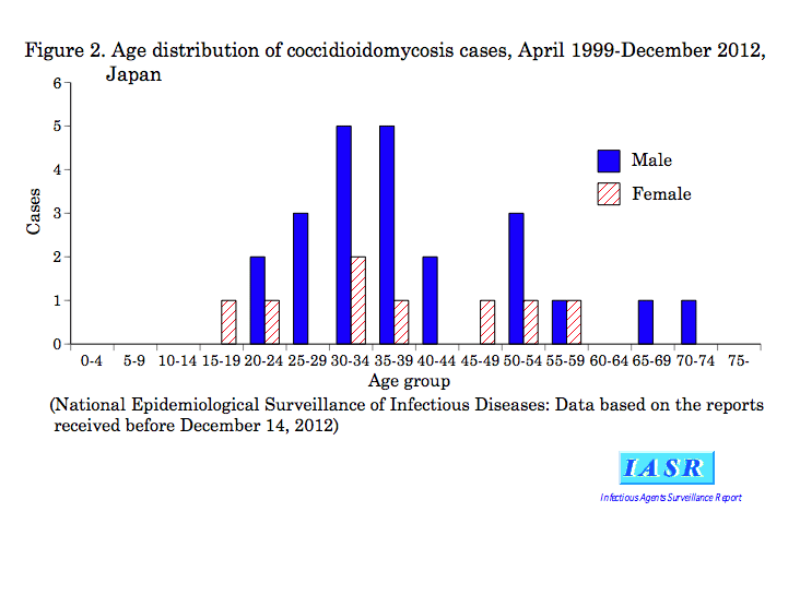
From April 1999 to December 2012, 31 cases were reported (Table 1 & Fig. 1); 23 cases were male and 8 were female, and those in their 30’s were the most frequent (13 cases) (Fig. 2). Infection of 27 cases among the total 31 cases was suspected to have occurred in the United States of America (14 in Arizona State).
Another member of the imported mycosis, histoplasmosis, is increasing in number though its frequency still remains in the single digits (see p. 3 of this issue).
Cryptococcus can infect healthy people. Once the pathogen invades the central nervous system, it may cause fatal consequence. According to the clinical laboratory data compiled for year 2011 within the framework of Japan Nosocomial Infections Surveillance (JANIS) sponsored by the Ministry of Health Labour and Welfare (MHLW), Cryptococcus occupied 2.8% of all the pathogens isolated from cerebrospinal fluids of encephalomeningitis patients (including both immunologically normal and compromised cases). The frequency was comparable to those of pneumococci, enterococci, and Escherichia coli.
Mycoses most frequently observed among immunocompromised patients are opportunistic infections by genus Candida and genus Aspergillus. While in clinical setting, candidiasis was preeminently frequent (Horn DL, et al., Clin Infect Dis. 48: 1695-1703, 2009), in the autopsy data from the Annual of Pathological Autopsy Cases in Japan, candidiasis and aspergillosis were equally frequent (2% of all the autopsied cases, respectively) (Kume H, et al. Med Mycol J 52: 117-127, 2011). It is probably because aspergillosis was more difficult to treat and more fatal than candidiasis.
Diagnosis and laboratory examinations
Isolation and identification of pathogenic fungi usually depend on morphological and biochemical characters, but for definitive identification, genetic methods are often used. Laboratory handling of pathogenic fungi requires special attention, as the fungal spores, particularly those of Coccidioides spp., which are dispersed from colonies developing on agar plates, may cause laboratory infection. Culture of Coccidioides spp. requires the biosafety level 3 facilities (BSL3) (see p. 3 of this issue). Therefore, whenever coccidioidomycosis is suspected, such as, from patient’s overseas travel history or residential place abroad, it is preferable to ask National Institute of Infectious Diseases or Medical Mycology Research Center, Chiba University, for advice on laboratory diagnosis.
Even when fungi cannot be isolated, diagnosis is possible based on the specific patho-histological characteristics of different mycoses.
As auxiliary methods, antigen detection can be used. Highly reliable are detection of glucuronoxylomannan antigen of genus Cryptococcus from sera or cerebrospinal fluids. Detection of galactomannan antigen of genus Aspergillus in sera is highly sensitive for patients with hematologic malignancies. Increase of antibody titer over clinical course will be an additional infection indicator.
Therapy of mycosis and development of drug resistance
Owing to availability of new generation antifungal drugs that effectively control the systemic mycoses (see p. 4 of this issue) and based on accumulation of clinical experiences, the standard treatment protocol has now been established. Even under the standard regimen, administration of antifungal agents may last from months to years, which increases the risk of appearance of drug resistant fungi. Actually, among fungi causing systemic mycosis, genetic mutations responsible for the drug resistance, though rare, have been reported, such as azole antifungal drug resistance mutations among Candida and Aspergillus (Tashiro M, et al., Antimicrob Agents Chemother. 56: 4870-4875, 2012) and echinocandin antifungal drug resistance mutation among Candida (Inui S, et al., J Jpn Assoc Infect Dis 85: 49-53, 2011).
Mycosis of public health importance
Other mycoses of public health importance are Cryptococcus gattii, a mycosis with high fatality, which is increasing in the Northern American Continent (see p. 4 of this issue) and Trychophyton tonsurans causing tinea capitis (see p. 5 of this issue) that is increasing among young adults in Japan. The issues are under investigation by research groups supported by the MHLW so as to establish appropriate countermeasures.
Note: Coccidioides immitis is regulated by Infectious Diseases Control Law as group 3 pathogen. When it is isolated, it should be reported to the MHLW within 7 days and when the isolates are to be transported it should be notified to Public Safety Commission.