Jpn. J. Infect. Dis., 55, 128-130, 2002
To see a printable version of the article in the Adobe file format, crick this [PDF] link.
Laboratory and Epideimiology Communications
Genotyping of Encephalitozoon cuniculi Isolates Found in Hokkaido
Koji Furuya*
Hokkaido Institute of Public Health, Kita-19, Nishi-12, Kita-ku, Sapporo 060-0819
Communicated by Kazuo Kato
(Accepted August 16, 2002)
Encephalitozoon cuniculi is an obligate intracellular protozoan that infects a wide range of mammalian hosts such as rabbits, rodents, dogs, other canids, and primates, including humans (1). Two reports have described the isolation of Encephalitozoon based on clinical materials obtained in Hokkaido (2,3). In 1995, Furuya et al. (2) reported Encephalitozoon-like organisms, describing the isolation of a microsporidian parasite from a human liver lesion surgically excised from a patient with alveolar hydatid disease. The organisms were identified by Nagano et al. as E. cuniculi (4). In 2001, Furuya et al. (3) also reported the isolation of Encephalitozoon organisms from a rabbit with neurological impairment at a municipal zoo in Hokkaido. They were also identified as E. cuniculi by means of polymerase chain reaction (PCR) and direct DNA sequencing. The former isolate was coded as 9504HF and the latter as 2008FF.
Recent E. cuniculi isolates from human and animal materials have been divided into three types, genotype I, genotype II, and genotype III, based on the number of 5'-GTTT-3' repeats in the internal transcribed spacer (ITS) of the ribosomal RNA genes (5,6). Genotype I had three tetranucleotide repeats, genotype II two repeats, and genotype III four repeats. There was a close relationship between genotype and host specificity. Genotype I was isolated from rabbits, genotype II from mice and blue foxes, and genotype III from domestic dogs (7). Genotypes I and III, but not type II, have been identified in human immunodeficiency virus (HIV)-infected patients. The author, therefore, typed the above two Hokkaido isolates by PCR and direct DNA sequencing.
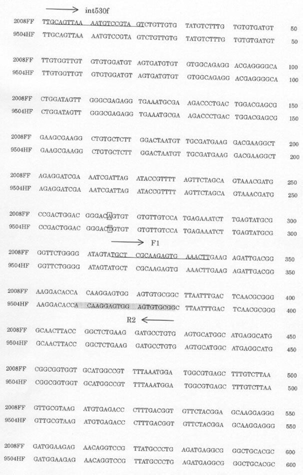
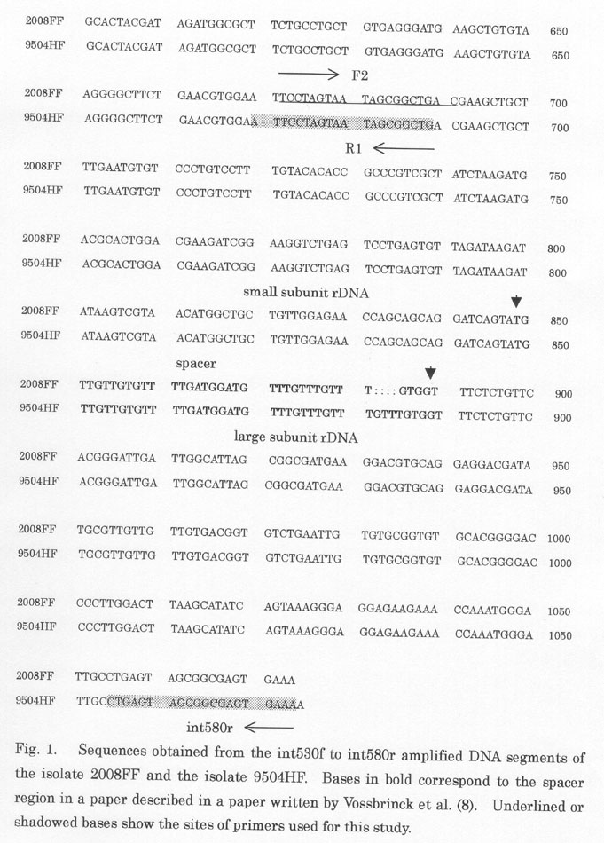
DNA samples were as described in a previous paper (3). PCR was carried out using the primer set of 5'-TGCAGTTAAAATGTCCGTAGT-3' (int530f) and 5'-TTTCACTCGCCGCTACTCAG-3' (int580r), which was originally designed by Didier et al. (5). A product of approximately 1,000 base pairs (bp) amplified with the primer set by PCR includes a large portion of the small subunit rRNA gene, the entire ITS, and a small portion of the large subunit rRNA gene (5,8). The nucleotide sequences of the both strands of the PCR products were determined using an ABI-PRISM BigDye terminator cycle sequencing ready reaction kit (Applied Biosystem, Foster City, Calif., USA) employing an ABI-PRISM 377XL DNA sequencer (Applied Biosystems). Four additional primers, F1 (5'-TGCTCGCAAGAGTGAAACTT-3'), F2 (5'-TCCTAGTAATAGCGGCTGAC-3'), R1 (5'-CAGCCGCTATTACTAGGAAT-3'), and R2 (5'-CCGCACACTCCACTCCTTGT-3') were used. Figure 1 (Fig. 1-Continued) shows sequences of the amplified DNA segments (1,070 bp for the isolate 2008FF and 1,075 bp for the isolate 9504HF). The sites of each primer are indicated; the small and large subunit rRNA gene regions with the ITS region are also shown. The isolates 2008FF and the 9504HF were identical except for one base-conversion and four nucleotides missing in 2008FF relative to 9504HF. One nucleotide difference was seen at position 266 (5'-CGGGACA(orT)GTG-3': A for the isolate 2008FF and T for the isolate 9504HF) in the 3' end of the small subunit rRNA gene as described before (3). Regarding this base-conversion, Xiao et al. (6) have recently reported that genotype I had A (5'-CGGGACAGTG-3') whereas genotype II and III had T (5'-CGGGACTGTG-3'). The sequence 5'-GTTT-3' in the ITS region was repeated three times in 2008FF and four times in the amplified 9504HF segment. The present results clearly suggested that the isolates 2008FF and 9504HF respectively belonged to genotypes I and III.
Human isolates of E. cuniculi have been identified as genotype I in Europe (9,10) and as genotype III in the Western Hemisphere (11,12). Accumulating evidence indicates that human infections with E. cuniucli result from animal sources. As described above, the isolate 2008FF (genotype I) was isolated from a diseased rabbit in the municipal zoo's colony where an outbreak of encephalitozoonosis occurred (3). Many pet rabbits kept in households have recently been found to be Eencephalitozoon antibody-positive (unpublished data). These observations suggest that E. cuniculi genotype I infections were common among rabbits in Hokkaido and that exposure to animal excrements is a potential cause of human infections. As for genotype III, represented by the isolate 9504HF, dogs may have been the source of infection to humans, because dogs and foxes are both definitive hosts in the life cycle of Echinococcus in Hokkaido (13) and 20 of 75 (26.7%) patients with alveolar hydatid disease in Hokkaido were dog owners (Sato, N., Hokkaido University Hospital, personal communication).
REFERENCES
*Corresponding author: Tel: +81-11-747-2739, Fax: +81-11-736-9476, E-mail: furuya@iph.pref.hokkaido.jp
Go to JJID Homepage Go to JJID Contents 55 (4)