Jpn. J. Infect. Dis., 56, 221-222, 2003
To see a printable version of the article in the Adobe file format, click this [PDF] link.
Laboratory and Epidemiology Communications
Atypical Proteinase K-Resistant Prion Protein (PrPres) Observed in an Apparently Healthy 23-Month-Old Holstein Steer
Yoshio Yamakawa*, KenŐichi Hagiwara, Kyoko Nohtomi, Yuko Nakamura, Masahiro Nishizima ,Yoshimi Higuchi1, Yuko Sato1, Tetsutaro Sata1 and the Expert Committee for BSE Diagnosis, Ministry of Health, Labour and Welfare of Japan2
Department of Biochemistry & Cell Biology and 1Department of Pathology, National Institute of Infectious Diseases, Tokyo 162-8640 and 2Miistry of Health, Labour and Welfare, Tokyo 100-8916
Communicated by Tetsutaro Sata
(Accepted December 2, 2003)
*Corresponding author: Mailing address: Department of Biochemistry and Cell Biology, National Institute of Infectious Diseases, Toyama 1-23-1, Shinjuku-ku, Tokyo 162-8640, Japan. Tel: +81-3-5285-1111, Fax: +81-3-5285-1157, E-mail: yamakawa@nih.go.jp
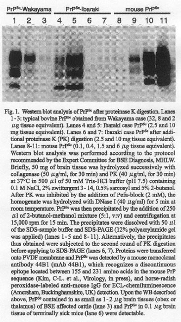
Since October 18, 2001, 'bovine spongiform encephalopathy (BSE) examination for all cattle slaughtered at abattoirs in the country' has been mandated in Japan by the Ministry of Health, Labour and Welfare (MHLW). 'Plateria' ELISA-kit (Bio-Rad Laboratories, Hercules, Calif., USA) is routinely used at abattoirs for detecting proteinase K (PK)-resistant prion protein (PrPSc) in the obex region. Samples positive according to the ELISA screening are further subjected to Western blot (WB) and histologic and immunohistochemical examination (IHC) at the National Institute of Infectious Diseases (NIID) or Obihiro University. If PrPSc is detected either by WB or by IHC, the cattle are diagnosed as BSE. The diagnosis is approved by the Expert Committee for BSE Diagnosis, MHLW. From October 18, 2001 to September 30, 2003, approximately 2.5 million cattle were screened at abattoirs. A hundred and ten specimens positive according to ELISA were subjected to WB/IHC. Seven showed positive by both WB and IHC, all exhibiting the typical electrophoretic profile of a high content of the di-glycosylated molecular form of PrPSc (1-3) and the distinctive granular deposition of PrPSc in neuronal cells and neuropil of the dorsal nucleus of vagus.
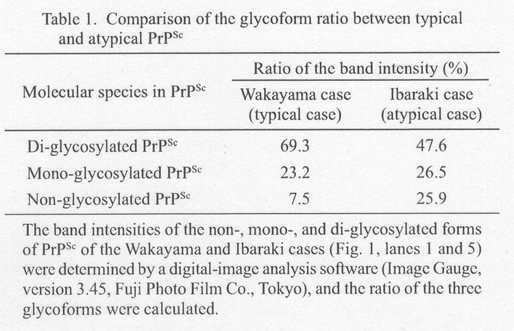
An ELISA-positive specimen from a 23 month-old Holstein steer slaughtered on September 29, 2003, in Ibaraki Prefecture (Ibaraki case) was sent to the NIID for confirmation. The animal was reportedly healthy before slaughter. The OD titer in ELISA was slightly higher than the 'cut-off' level given by the manufacturer. The histology showed no spongiform changes and IHC revealed no signal of PrPSc accumulation typical for BSE. However, WB analysis of the homogenate that was prepared from the obex region and used for ELISA revealed a small amount of PrPSc with an electrophoretic profile different from that of typical BSE-associated PrPSc (1-3). The characteristics were (i) low content of the di-glycosylated molecular form of PrPSc, (ii) a faster migration of the non-glycosylated form of PrPSc on SDS-PAGE, and (iii) less resistance against PK digestion as compared with an authentic PrPSc specimen derived from an 83-month-old Holstein (Wakayama case) (Fig. 1). Table 1 summarizes the relative amounts of three distinctive glycoforms (di-, mono, non-glycosylated) of PrPSc calculated by densitometric analysis of the blot shown in Fig. 1. As 2.5 mg wet weight obex-equivalent homogenate of the Ibaraki case (Fig. 1, lane 4) gave slightly stronger band intensities of PrPSc than an 8 mg wet weight obex-equivqlent homogenate of a typical BSE-affected Wakayama case (Fig. 1, lane 2), the amount of PrPSc accumulated in the Ibaraki case was calculated to be 1/500 - 1/1000 of the Wakayama case. In the Ibaraki case, the PrPSc bands were not detectable in the homogenates of the proximal surrounding region of the obex. These findings were consistent with the low OD value in ELISA, i.e., 0.2 - 0.3 for the Ibaraki case versus over 3.0 for the Wakayama case. The DNA sequence of the PrP coding region of the Ibaraki case was the same as that appearing in the database (GenBank accession number: AJ298878). More recently, we encountered another case that resembled the Ibaraki case. It was a 21-month-old Holstein steer from Hiroshima Prefecture. WB showed typical BSE-specific PrPSc deposition though IHC did not detect positive signals of PrPSc (data not shown).
Though the clinical onset of BSE is usually at around 5 years of age or later, a 20-month-old case showing the clinical signs has been reported (4). Variant forms of BSE similar to our cases, i.e., with atypical histopathological and/or biochemical phenotype, have been recently reported in Italy (5) and in France (6). Such variant BSE was not associated with mutations in the prion protein (PrP) coding region as in our case (5,6).
The Ministry of Agriculture, Forestry and Fisheries of Japan (MAFF) announced a ban of feeding ruminants with meat bone meal (MBM) on September 18, 2001, and a complete ban was made on October 15 of the same year. According to the recent MAFF report, the previous seven cases of BSE in Japan were cattle born in 1995 - 1996 and possibly fed with cross-contaminated feed. However, the two cattle in this report were born after the complete ban. Whether contaminated MBM was implicated in the present cases remains to be investigated.
REFERENCES
Go to JJID Homepage Go to JJID 56 (5, 6) Contents