Jpn. J. Infect. Dis., 55, 99-100, 2002
Laboratory and Epidemiology Communications
Amplification of Irrelevant Sequence from Bacillus subtilis Using a Primer Set Designed for Detection of the pag Gene of Bacillus anthracis
Osamu Fujita, Satoshi Inoue, Masashi Tatsumi, Tsuneo Kamiyama, Shoichi Akaishi1, Tomoko Ootani1, Tsuneaki Kawai1, Takashi Hirochi1, Yumiko Sakamoto1, Kazumichi Tamura2, Haruo Watanabe2 and Akio Yamada*
Department of Veterinary Science and 2Department of Bacteriology I, National Institute of Infectious Diseases, 1-23-1 Toyama, Shinjuku-ku, Tokyo 162-8640 and 1Sapporo City Institute of Public Heath, Kikusui 9-1-5-22, Siroishi-ku, Sapporo, Hokkaido 003-8505
Communicated by Kazuo Kato
(Accepted July 26, 2002)
Anthrax is one of the major bacterial zoonoses caused by spore-forming
Gram-positive rods, Bacillus anthracis. After the bioterrorist
attack in the USA in September 2001, the probability of anthrax
attacks has increased worldwide, and the number of hoaxes involving
"white powder" has increased in Japan as well. Since
rapid and precise diagnosis is the most powerful measure to counter
bioterrorism, it is of particularly importance to have a diagnostic
tool to detect anthrax. Polymerase chain reaction (PCR) is regarded
as one of such diagnostic method for the detection of anthrax,
although definitive diagnosis can only be made when other methods
support the PCR results. The PCR method recommended by the World
Health Organization (WHO) is widely accepted in Japan as the standard
approach. A presumptive diagnosis of anthrax is made if bands
with predicted sizes are detected after PCR with primer sets targeted
to pag and cap genes residing on pXO1 and pXO2 plasmids,
respectively (1).
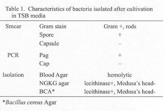
On March 27, 2002, "white powder" was found disseminated inside a public restroom located in Sapporo City. The powder was subjected to microbiologic tests and PCR at the Sapporo City Institute of Public Heath directly and after cultivation in TSB medium. Although no bacterium was found when the sample was directly examined, Gram-positive, spore-forming bacilli were detected after cultivation in TSB medium (Table 1). Since the bacteria were motile and hemolytic, the possibility that isolated bacteria were B. anthracis was ruled out; however, the PCR with primers designed for detection of the pag gene resulted in amplification of a 596 bp fragment (Fig. 1). The presence of the PCR amplicon of this size usually indicates that the sample may contain bacteria harboring a pXO1 pathogenic plasmid. We have thus attempted to determine whether or not the bacteria were indeed B. anthracis. First, microplate hybridization (kindly provided by Dr. Matsunaga of Wakunaga Pharmaceutical Co., Ltd., Hiroshima) was employed for detection of the Ba813 chromosomal gene that was thought to be anthrax-specific (2). The results revealed no significant signals, suggesting that the bacteria did not have sequences homologous to Ba813. Second, real-time PCR was performed using a LightCycler - Bacillus anthracis Detection Kit (Roche Diagnostics, Tokyo). No anthrax-specific signals were detected when the sample was tested for the presence of pag and cap genes. We have then sequenced the amplicon after cloning into pCR2.1 plasmid. The nucleotide sequence showed substantial homology with B. subtilis ATP-dependent deoxyribonuclease, but no homology with any of known B. anthracis genes was determined. An increase of the annealing temperature had no effect on the production of these bands when the sample was subjected to PCR using the same pag primer set (Fig. 1). The results of biochemical tests indicated that the bacteria isolated belonged to B. subtilis group (data not shown). These findings indicate that the PCR protocol recommended by WHO is not sufficiently specific, unless the internal nucleotide sequence is determined, since there remains a possibility that a bacterium exists, the genome of which contains sequence(s) amplifiable by the primers. Moreover, the size of amplified fragments is indistinguishable from those amplified from B. anthracis.
This article appeared in the Infectious Agents Surveillance Report, vol. 23, no. 7, 2002 in Japanese.
REFERENCES
Go to JJID Homepage Go to JJID Contents 55 (3)