![]()
The Topic of This Month Vol. 33, No. 10 (No. 392)
Mycoplasmal pneumonia as of September 2012, Japan
(IASR 33: 261-262, October 2012)
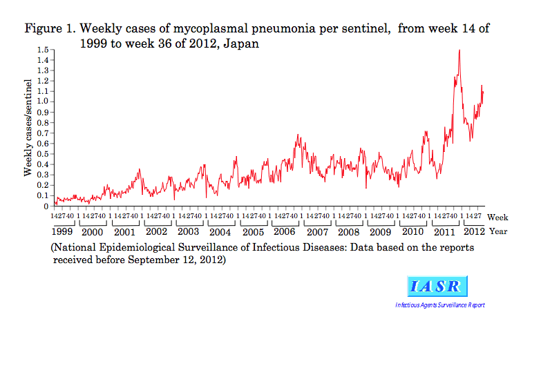
Mycoplasmal pneumonia is a category V infectious disease in the National Epidemiological Surveillance of Infectious Diseases (NESID) under the Law Concerning the Prevention of Infectious Diseases and Medical Care for Patients of Infections (the Infectious Diseases Control Law) enforced on April 1, 1999. Sentinel hospitals* regularly report the weekly number of mycoplasmal pneumonia patients (total of outpatients and inpatients). In addition to isolation of Mycoplasma pneumoniae or detection of serum antibody to M. pneumoniae, detection of M. pneumoniae genome by PCR or LAMP has been included in the notification criteria (http://www.mhlw.go.jp/bunya/kenkou/kekkaku-kansenshou11/01-05-38.html) since its modification in April 2011. Recently, the reported number of mycoplasmal pneumonia patients is increasing (Fig. 1).
*There are about 500 sentinel hospitals in Japan, which are selected from those equipped with departments of pediatrics and internal medicine and with more than 300 beds.
Periodicity of mycoplasmal pneumonia epidemics and the epidemic that started in 2011: Periodicity of mycoplasmal pneumonia epidemic at a 3-8 year interval has been observed worldwide. It is presumably brought about by the herd immunity vs. pathogen interaction but its exact mechanism is unknown. Seasonally, mycoplasmal pneumonia is prevalent from autumn to winter, and occasionally also in early summer (Fig. 1).
In Japan, the large-scale atypical pneumonia epidemics occurred at four-year intervals from the late 1970s through the 1980s coinciding Olympic years. Under the former NESID system (July 1981-March 1999), number of “clinically-diagnosed atypical pneumonia” cases peaked in 1984 and 1988 (IASR 28: 31-32, 2007) and it was probably epidemics of mycoplasmal pneumonia, because major cause of atypical pneumonia is M. pneumoniae. Though almost absent since 1990s (except the epidemic in 2006), mycoplasmal pneumonia cases increased in autumn of 2010 (Fig. 1). In 2011, number of the reports increased from summer and reached its peak in winter, which was more than twice as high as the past peaks in 2006 and 2010. The number of the reports per week in 2012 is continuously higher than in 2011. Since 2010, mycoplasmal pneumonia epidemic is found worldwide, such as, in United Kingdom, France, Northern European countries and Israel.
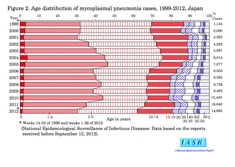
Age and geographical distribution of mycoplasmal pneumonia patients: Children aged 1-14 years occupied 80% of the patients. Among them, since 2011, age group 10-14 years increased and that below 4 years decreased in proportion (Fig. 2). As such age group shift has been observed in the past (IASR 28: 31-32, 2007), the present resurgence of mycoplasmal pneumonia may not be related to the age shift.
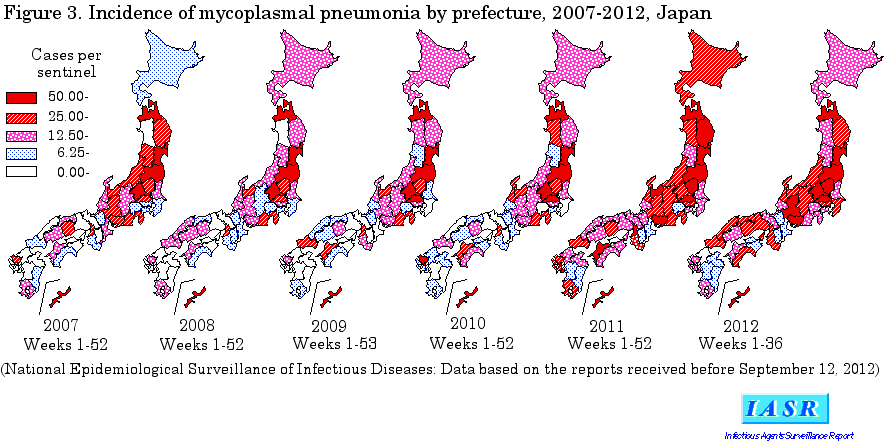
Regionally, since 2007, Aomori, Miyagi, Fukushima, Gunma, Saitama and Okinawa sentinel points have reported larger number of mycoplasmal pneumonia cases, and since 2010, Iwate, Tochigi, Toyama, Aichi, Gifu, Osaka, Ehime and Saga also do so (Fig. 3). Since 2011, other prefectures with fewer cases started to report larger number of cases than before.
Mycoplasma pneumoniae: Among species of Mycoplasma of human origin, clear pathogenicity has been found only in Mycoplasma pneumoniae. M. pneumoniae belongs genus Mycoplasma of class Mollicutes. Its genome is 800kb and the smallest among organisms that grow in artificial media. It is entirely devoid of peptide glycan cell wall, and β-lactam antibiotics are ineffective. It is rod-shaped and 0.3 × 2 µm in size. It has a cytoplasmic protrusion on one end of the body; it is an organelle used for adherence to the surface of respiratory epithelial cells thus contributing to the bacteria’s pathogenicity (see p. 263 of this issue). On its surface clustered is a large number of cytadhesin protein P1 (molecular weight 170kDa). P1 protein is polymorphic, which allows classification of M. pneumoniae into type 1 and 2 and their subtypes by genome sequencing. So far, type 1, 2, 2a, 2b and 2c have been identified among the Japanese clinical isolates. Though the type does not affect the pathogenicity, the frequency of the prevalent types is variable from year to year and from region to region. Recently, multiple-locus variable-number tandem repeat analysis (MLVA) is used for epidemiological investigation in the US and European countries. So far, more than 30 MLVA types have been reported.
Increase of macrolide resistance: Macrolide antibiotics are used for treatment of mycoplasmal pneumonia. However, since 2000 when the macrolide-resistant M. pneumoniae was first reported in Japan, macrolide-resistant strains are found continuously increasing in Asia and nearby regions (IASR 32: 337-339, 2011). Now, more than 50% of clinical isolates in Japan are estimated to be macrolide resistant (see pp. 264, 265 & 267 of this issue). However, European countries experiencing mycoplasmal pneumonia epidemic similarly as Japan report the macrolide resistance rate below 10%. Macrolide-resistant strains are more often isolated from pediatric patients rather than adults. Macrolide resistance itself does not significantly affect the sequel as most cases recover without chemotherapy. When the patients received chemotherapy, feverish phase may prolong in case of macrolide-resistant M. pneumoniae infection than in case of macrolide-susceptible one (IASR 28: 41-42, 2007 and see p. 266 of this issue). Macrolide-resistant M. pneumoniae infection can be effectively treated with quinolone and tetracycline antibiotics, and no resistant clinical isolates have been reported in the world including Japan. However, their use for children should be limited to really serious cases on account of their potential side effects (see p. 268 of this issue).
Laboratory diagnosis of mycoplasmal pneumonia: Culture, serodiagnosis and gene amplification methods are available. Isolation of M. pneumoniae is the most reliable, but 1-4 weeks are required for obtaining the final data. PA, EIA and other serodiagnostic kits are preferentially used in clinical settings for the rapid test. However, it is useful only when the paired serum antibody titers of the patients were obtained. The current most reliable rapid diagnosis is PCR, LAMP and other gene amplification methods (see p. 268 of this issue). LAMP method has been covered by the health insurance since October 2011.
Final Comments: The progressing mycoplasmal pneumonia epidemic in Japan (http://www.niid.go.jp/niid/ja/10/2096-weeklygraph/1659-18myco.html) necessitates continued surveillance of patients and continued monitoring of drug-resistance and pathogenicity of the bacterial isolates.